Version 2.1
Download: SetupCIMicro2.1.exe (CIMicro needs .NET Framework 4.6.1 or higher)
Copyright © CellularImaging – Core Facilty AMC, Ron Hoebe, 2015-2019
CI Micro can show and analyze images (Single, Stacks and Time Series) from LAS-X Files; LIF, XLEF or TIF.
This program was developed primary for quickly analyzing time-lapse experiments of cell migration, invasion and wound healing assays. Time-lapse experiments saved in Leica’s LIF or (auto-saved) XLEF files can be opened with no delay in loading time. Interactive measurements can be done by the area or counting tool and are save together with experimental data. Extra options are Movie Export, 3D features and support for TIFF files.
Image Data and Meta Data is only read when needed. This makes access to large data sets very quick. Original Files are opened as read-only and are never altered.
Main Form | Viewer Tools | Interactive Count | Interactive Area | Special | Known issues | Work in progress | Release Notes
Main Form: Choose a LIF/XLEF file or Export Folder. Select an image, single, stack or image time series in the treeview to display.
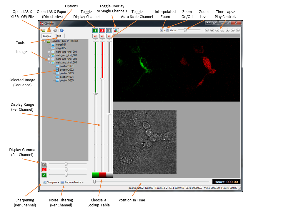
Viewer tools:
- Select a region of interest (ROI) and double click this ROI to Crop. Double click the cropped image to cancel cropping.
- Use the arrow keys for stepping trough an image sequence (CTRL and SHIFT make bigger steps).
- Stacks can be viewed as single slices, maximum intensity projection or maximum variance projection.
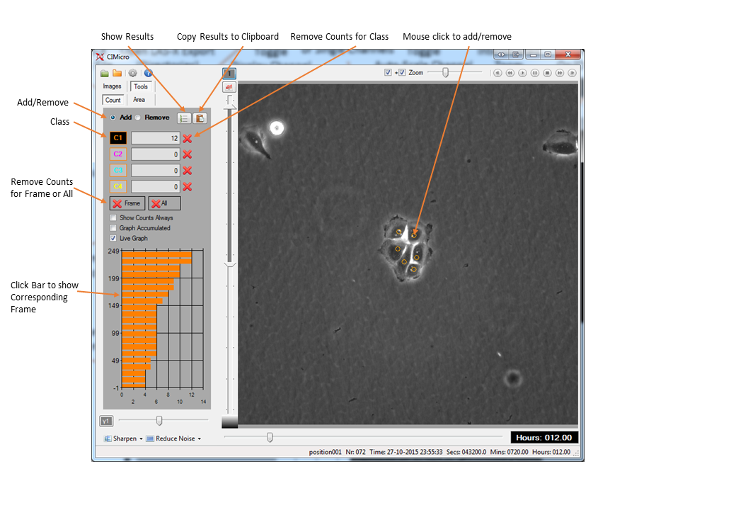
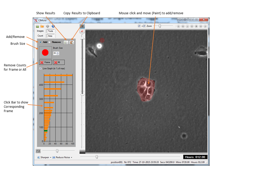
Interactive tools:
Count: Use one of the four Classes to count events or features (in this example; the Number of Cells)
Area: Paint with a Brush to Measure an Area (In this example; the Area of one Clone). Use right click to Flood Fill an Area.
Special Features:
- CIMicro can read Autosave XLEF/LOF data even when the software crashes (like with a power failure). The timestamps of a time-series are lost and set to 1 hour between images at default (This can be changed by the setting: LostTimeInterval)
- XLEF: Data is stored in a \db folder in the \metadata folder of the original data, LIF: Data is stored in a \db_LIF_Filename folder in the same folder as the original LIF file (copy them together if you move your data to keep settings and analysis)
- Export Time Series to Movie (TAB not shown in screenshots)
- 3D Viewer
Not shown in screenshots (only visible with stacks).
Keys and Mouse functions:
| Zoom: | PageUp, PageDown , MouseWheel |
| Rotation Speed: | Left, Right, Up, Down |
| Stop Rotation: | Spacebar, Esc |
| Light On: | L |
| Light Off: | K |
| Transparency: (when Light is off) | T, Y |
| Red Color (or Light): | R, E |
| Green Color (or Light): | G, F |
| Blue Color (or Light): | B, V |
| All Colors (or Light): | Add, Subtract (+, – on numkeypad) |
| Increase Z scale | X |
| Decrease Z scale: | Z |
| Rotate | Hold Left Mouse Button and Drag |
| Move | Hold Right Mouse Button and Drag |
- Remark: When light is on (the default) the keys (R,E,G,F,B,V) control the ambient light parameters, if light is off the keys control the color parameters.
- Remark: When the 3D Picture seems overexposed at start. Reduce the overall light by Add, Subtract (+, – on numkeypad)
- Bug: When the 3D viewer crashes. Restart the complete program. (Looks like a locked temporary file problem)
Known limitations and issues:
- Maximum of 6 Channels
- Export to Movie is stable but a bit slow.
- Beta version, so there are probably still a few bugs!
- Memory limit (everything up to 2048×2048 pixels and up to 6 channels and 100 3D slices works fine, larger images with multiple channels can give memory errors)
Work in progress:
- Better Export (to TIFF does work, right click an image in the Treeview and select “Export Current Image”)
- Plugins (Matlab and Huygens)
- Maybe an additional 64 bits version (the program is now a 32 bits version, running on 32-bits and 64 bits Windows 7 and higher using the .NET Framework 4.6.1)
Release Notes:
- Version 2.1
- Fixed: Small Bug
- Version 2.0
- TIFF Export file contain correct pixel size
- Fixed: Bugs
- Version 1.4
- New: 3D Viewer (as is, final version)
- New: TIFF file support by selecting folder of TIFF Files (8 and 16 bits greyscale (1 channel) and 24 or 48 bits color (3 channels))
- New: ROI Size (popup menu, right click picture with a selection)
- Fixed: Image loading speed speed
- Fixed: Bugs
- Version 1.3
- New: LIF File support
- New: Image Stack Support, with Max Intensity projection and Max Variance projection
- New: Area Tool; Brush color and transparency
- Fixed: Image levels
- Fixed: Bugs
- Version 1.2
- New: Movie Export of Time Series
- New: LostTimeStamp (experiment crash problem)
- Fixed: Bugs
- Version 1.1
- New: Area Tool (This is mainly used for the scratch (wound healing) assay)
- New: Count Tool
- Fixed: Bugs
- Version 1.0
- Initial Version
Copyright © CellularImaging, 2015-2019
Core Facilty, AmsterdamUMC, AMC, Ron Hoebe
r.a.hoebe@amsterdamumc.nl
http://software.cellularimaging.nl
http://cellularimaging.nl